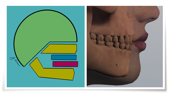
Classification of Face
It is not sufficient to categorize orthodontic malocclusions on the basis of a classification of the teeth alone. The relationship with other craniofacial structures must also be taken into consideration.
Class I:
Maxillary-Mandibular Dental Protraction - Teeth (Dentition)
This is an example of a dental malocclusion that may require the removal of teeth for correction.
Maxillary-Mandibular Dental Retraction - Teeth (Dentition)
This is an example of a dental malocclusion that may be treated with expansion rather than removing teeth.
Class II:
Maxillary Dental Protraction - Teeth (Dentition)
This is an example of a dental malocclusion that may require the removal of teeth for correction.
Mandibular Retrognathism - Lower jaw
The lower jaw has not grown as much as the upper jaw. This example of a Class II malocclusion demonstrates the need for early growth guidance.
Maxillary Dental Protraction of the Teeth combined with Mandibular Retrognathism of the Lower Jaw
These Class II malocclusions are more difficult to treat due to the skeletal disharmony and may require orthognathic surgery in conjunction with orthodontic treatment.
Class III:
Mandibular Dental Protraction - Lower Teeth (Dentition)
The lower teeth are too far in front of the upper teeth. This malocclusion is treated with orthodontic procedures which may require the extraction of teeth due to the dental protrusion.
Mandibular Prognathism - Lower Jaw
The lower jaw has outgrown the upper jaw. This malocclusion is more difficult to treat due to the skeletal disharmony and may require orthognathic surgery in conjunction with orthodontic treatment.
Severe Skeletal Class II
Skeletal Open Bite
Generally, a set of cascading steps are often observed in the following order: 1) This condition is generally associated with a genetic downward and backward growth of the mandible at the chin (Bjork). In addition, this condition can be exacerbated by also genetically-controlled or environmentally induced enlargement of the Tonsils, Adenoids, Turbinates that humidify air, or Septum deviation shown in black, including asthma, allergies, sinusitis etc., act to often block the nasal and throat passage (nasal-pharyngeal obstruction). This results in the exacerbation of a chronic mouthbreathing pattern that can be habitual 2) Muscle activity is weak as the vertical dimension opens and the tongue lowers its natural position down from the palate 3) This results in Skeletal upper jaw (maxillary) constriction as the lower jaw is also positioned downward and backward 4) Biological, physiologic eruption of the posterior upper and lower molar segments additionally develops with the mouthbreathing resulting in an a skeletal anterior open bite where the strong tongue is also anteriorly positioned 800-1,000X/day during swallowing, speaking and eating to try to seal the anterior opening between the front teeth to contain the saliva with the oral cavity.
This condition is a medical one and often requires the first steps of ENT examination and Allergist evaluation to determine the cause (etiology) of the condition for a proper diagnosis that is a key factor. Once the etiology is determined and controlled the upper jaw (maxilla) may be expanded and the posterior first molar and also premolar regions may be re-intruded to close the open bite. Due to the strong tongue muscle function positioned anteriorly, this often needs to be controlled by 12 tongue stars bonded to the palatal of the upper incisors and lingual of the lower incisors (or 12 Aligner Tongue Trainers, ATTs) during treatment. It is important to note, that by controlling or ideally eliminating the etiology, the prognosis for the correction of the skeletal open bite improves to become more stable in retention.
Skeletal Deep Bite
This condition is generally associated with 1) a genetic upward and forward growth direction of the chin of the lower jaw, or mandible (Bjork). In addition, the condition is often associate with 2) muscle hyperactivity and 3) intrusion of the lower first molars and premolars (buccal segments) that are also in-line with the strong masseter-medial pterygoid elevator muscles shown in black diagonal lines. This results in a deep curve of Spee when the lower arch is viewed from the side because of the skeletal restriction of lower jaw (mandibular) buccal segment eruption early in development.
This condition commonly requires the use of Anterior bite ramps called Biturbos bonded to the upper front incisors to reduce muscle hyperactivity and allow the leveling of the deep curve of Spee. In addition, a new Anterior Intrusion Appliance (AIA) is indicated from the scientific evidence-based dated to be used to intrude the lower incisors. This is particularly true with thermoplastic aligner treatment to assist the aligner due to force loss after three days.
Skeletal Deep Bite with mandibular Retrognathism - lower jaw
This condition is usually treated with a JVBarre 4D for molar rotation with permanent Class II NiTi coils as a Class II corrector or advancer timed at the peak growth spurt. In adults surgical lower jaw advancement (mandibular) may be an option to balance the harmony of the profile.
Skeletal Deep Bite combined with Mandibular Retrognathism and Maxillary protraction of the teeth (dentition)
This condition is similar to the one above in 9) except the lower jaw is smaller or is positioned further back that may also be related to an obtuse cranial base angle shown.
The JVBarre 4D® is applied to the upper canines and molars with a fixed NiTi coil spring to keep the mandible forward and allow lower jaw joint, or mandibular condyle and fossa, to modify in growing children as the bite is re-opened. Dr. Voudouris has identified in fact 12 different contributions to the correction of Skeletal Deep Bite with Mandibular Retrognathism listed in the Supercorrection Prescription textbook.
The upper incisors are retracted in the final stage of treatment. An alternative may be a Herbst appliance carefully monitored for condylar resorption due to the chronic continuous advancement, or a removable Invisalign Mandibular Advance (MA)®, or removable Twin Block.
Skeletal Deep Bite combined with mandibular retraction of the incisors (or retroclination)
This condition is similar to the one above in 9) and related once again to a genetic upward and forward growth direction of the lower jaw (mandible) at the chin (Bjork) and muscle hyperactivity shown in diagonal black lines.
Due to the large size or forward position of the lower jaw (mandible), it is important not to move the retroclined lower incisors forward until the lower back molars and premolars (buccal segments) are differentially extruded to open the deep bite.
This opening of the bite positions the lower jaw downward and most importantly backward first to secondly, allow the alignment of the lower incisors. This prevents a Class III relationship with lower incisor protraction (please see 6) above).
This condition is shown in fact, in a patient here in our website and is classified as “Atypical” since skeletal deep bite is more commonly observed with a retrognathic mandible shown in 10) above or a more well-positioned upper jaw and well-positioned lower jaw (orthognathic) shown in 9) above.


